Electrical Charge Movement
In this lesson, we will describe how the electrical charge moves inside the heart.
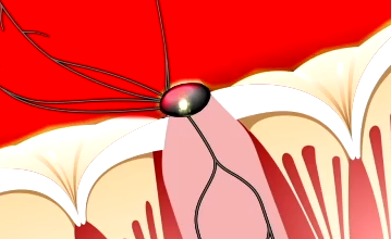
The video below shows a simplified version of the normal movement of the electrical charge through the normal heart: The electrical signal starts in the sino-atrial node (SA node). Note that it is located in the right atrium, near the top. This is important because in the next image, you can see the direction of depolarization through the atria.

We have used a thin yellow line to show the depolarization. The red shading indicates tissue that is currently depolarized (myocytes stay depolarized for a period of time). Remember that tissue that is currently depolarized cannot be depolarized again while it is in this state. Therefore, red shaded tissue is refractory to any new electrical signals that might arrive.

When the electrical signal reaches the atrio-ventricular node (AV node), it slows down a lot! The AV node becomes refractory, as indicated by the red shading for a brief time after it transmits a signal. This refractory period helps to limit the signals that go into the ventricles when there is a problem in the atria of having too many electrical signals (such as in atrial fibrillation or atrial flutter, 2 diagnoses you will learn about in future lessons).
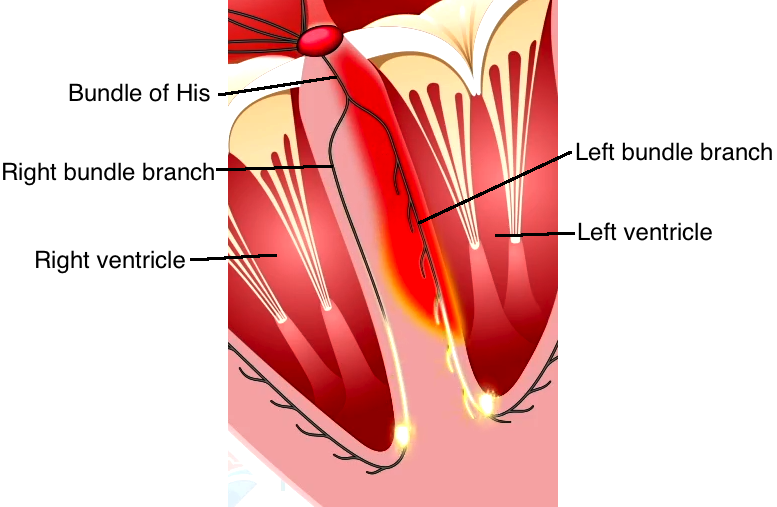
When the electrical signal exits the AV node, it enters the specialized conducting system call the bundle of His (the first part), the bundle branches (2 major divisions of left and right) and the little tiny branches. All these conducting cells are made of Purkinje fibers. They are special because they conduct the signal very fast. Notice how the signal travels down the Purkinje fibers well before the signal enters the myocardium to make it shade red.
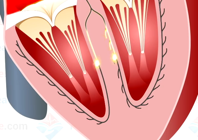
The initial part of depolarization of the ventricles is very important. Note that the septum is the first to depolarize and that it depolarizes starting on the left side of the heart (which is the right side of this diagram). Notice that there are branches on the left bundle branch, but not on the right bundle branch.
The purkinje fibers are located close to the endocardium. As a result, depolarization in the ventricles begins near the endocardium and travels toward the epicardium. This is important because you would not want the epicardial myocytes contracting first, on top of relaxed myocytes near the endocardium. This would not be efficient. It is important to pay attention to the direction of depolarization in the ventricles for later ECG concepts.

The ventricles are now in a state of depolarization for a short period of time. This is to provide time for mechanical contraction to occur. Electrically speaking, it occurs because the action potential has a plateau phase before repolarization (review lesson 2 on action potentials to see the plateau).

We have used the color blue to show repolarization. Notice that it is not very fast and most importantly, notice that it is in the opposite direction (epicardium to endocardium) compared to depolarization. This is important because mechanically, you would not want cells near the inside endocardium to start relaxing while the outside epicardial cells were still contracting. This would be very inefficient. The plateau phase of the action potential of the myocytes near the outside of the heart are very short, so these cells stay depolarized for the shortest period of time.