Rule: More P waves than QRS complexes = conduction block
If you have a P wave without a QRS after it, then the electrical signal that generated the P wave did not travel into the ventricles to generate a QRS complex. Therefore, the electrical signal was blocked.
The only way to have more P waves than QRS complexes is to have one or more P waves without a QRS after it. There are 2 situations in which you will see this:- The AV node is diseased and is not conducting the electrical signals from the SA node down to the ventricles.
- The AV node is normal, but there are too many P waves (more than 200 per minute) that can be conducted through the AV node, so there is a conduction block for normal physiological reasons.
Listing these situations above as ECG diagnoses, they are:- Second or third degree heart blocks
- Atrial flutter (with a physiological block)
Note that atrial fibrillation is not listed because there are no P waves with atrial fibrillation and first degree heart block is not listed because with
first degree block, there are an
equal number of P waves with QRS complexes.
If these terms are not familiar to you yet, don't stress. We will cover them in the Rhythm Diagnostic Criteria tutorial.Rule: P waves all the same size and shape = 1 origin for the P waves
This should make sense. If the P waves are all starting from the exact same location within the heart, then they will travel through the heart in the same way and create the same deflection every time on the ECG.Rule: P waves NOT all the same size and shape = 2 or more origins for the P waves
If you depolarize the atria from a different location, then the signal will travel through the atria in a different direction and have a different vector and therefore create a different waveform on the ECG. Below is atrial depolarization by a sinus, atrial, and junctional pacemaker. Each will create a unique wave of electricity through the atria and therefore create a uniquely shaped P wave.
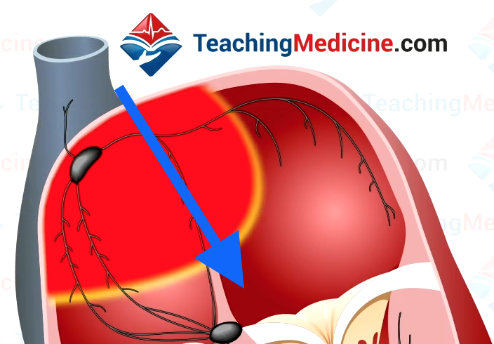
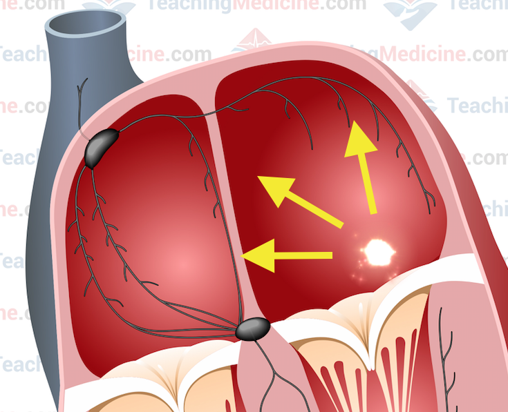
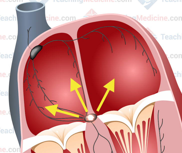
SA node beat Atrial beat Junctional beat
Sometimes we can see a mixture of pacemakers. For example, the SA node might be producing most of the heartbeats but then occasionally an extra beat from the atria or from the AV node might sneak in for a cameo.
Summary:- more P waves than QRS complexes = conduction block
- think 2nd or 3rd degree AV block
- think atrial flutter (or other fast atrial rhythms)
- P waves not all same size and shape = more than one origin for the P waves
- think about a combination of sinus, atrial or junctional beats
